Radial Nerve Compression Specialist

Are you an athlete or worker who participates in continual strenuous use of the arm and forearm? If so, you may be at risk of a condition known as radial nerve compression. Radial nerve compression causes deep pain in the forearm that can radiate to the fingers. Radial nerve compression specialist, Dr. James Mazzara provides diagnosis and both surgical and nonsurgical treatment options for patients in Manchester, South Windsor, Enfield, Glastonbury and surrounding Hartford communities who are experiencing symptoms of radial nerve compression. Contact Dr. Mazzara’s team today!
Radial Tunnel Syndrome & Posterior Interosseus Nerve Syndrome
What is Radial Nerve Compression at the Elbow?
One of the more difficult to make diagnoses to make in the upper extremity is distinguishing between radial tunnel syndrome and lateral epicondylitis. Both entities are caused by the same type of problem and may occur consurently in the same patient. The cause is generally related to repetitive and strenuous use of the arm.
The radial nerve is composed of the roots of C6-C8 which form the posterior cord of the brachial plexus. The nerve courses distally along the humerus to the lateral intermuscular septum is found. This is a potential site of compression. The radial nerve then bifurcates into a superficial sensory branch and a deep motor branch (the posterior interosseus nerve) that passes through the radial tunnel.
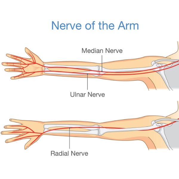
Click to Enlarge Image
The radial tunnel is composed of the radiohumeral joint and the superficial head of the supinator muscle where there is a fibrous band known as the arcade of Frohse.
What is Radial Tunnel Syndrome?
The radial tunnel syndrome results from dynamic compression of the posterior interosseus nerve in its course from the anterior capsule of the elbow joint proximally to the arcade of Frohse distally.
Symptoms include deep, dull proximal dorsal forearm ache, often with distal radiation. The pain is often described as a cramp. Night pain is common. Sensory loss over the dorsoradial aspect of the second metacarpal head suggests radial sensory branch involvement. Motor findings are usually absent.
On physical examination, symptoms are aggravated by the following:
- resisted supination and extension
- resisted extension in the metacarpophalangeal joint of the long finger with the wrist extended
- repetitive forearm pronation with the wrist flexed.
How to Diagnose Radial Tunnel Syndrome
EMG/NCS are not helpful in confirming the diagnosis but may be useful in identifying coexisting pathology.
Injections into the lateral epicondylar area can sometimes help differentiate radial tunnel syndrome from lateral epicondylitis.
What are the Treatment Options for Radial Tunnel Syndrome?
Conservative treatment is attempted in most cases. Efforts should be made to modify patient activity to avoid provocative positioning of the arm. Ergonomic evaluation should be completed to modify the offending task or job. Task that require elbow extension, forearm pronation and wrist flexion repetitively or for long periods of time contribute to the development of radial tunnel syndrome.
Initial treatment should include rest, stretching and splinting. Corticosteroid injections adjacent to, but not within, the nerve is an acceptable treatment option.
Do I Need Surgery for Radial Tunnel Syndrome?
Surgical intervention may be considered if the symptoms are not relieved by rest, activity modification, nonsteroidal anti-inflammatory medication, or a corticosteroid injection. Before considering surgery, precise localization of the pain to the radial tunnel must be confirmed.
Two surgical approaches are acceptable. First, an incision directly over the lateral epicondyle and supinator is considered when the surgeon is certain that the site of compression is the supinator at the Arcade of Frohse.
If doubt exists because of tenderness proximally over the radieal nerve, a more extensile approach is necessary. Care is taken to identify and release all potential sites of radial nerve compression.
Postoperative care involves a bulky long arm dressing to immobilize the elbow for one week. Gradual stretching and strengthening exercises are started at 1 week. Unlimited activities are permitted at 6-12 weeks depending on job requirements.
A gradual return to work program is favored for workers returning to more physically demanding work.
What is Posterior Interosseus Nerve Syndrome?
Posterior interosseus nerve syndrome is a compression neuropathy that presents with weakness or loss of function of the finger and thumb extensors. Conversely, radial tunnel syndrome is associated with pain and little, if any, weakness.
The symptoms may occur after an episode of strenuous arm and/or wrist use involving pronation and supination. Pain at the elbow may precede the weakness, which is usually rapid in progression and may be sequential.
Posterior interosseous nerve syndrome is associated with space occupying lesions such as ganglia. Other possible causes may include synovitis of the elbow joint, and compression by the radial recurrent artery. If compression is secondary to a space occupying lesion, onset may be gradual.
Posterior interossues nerve compression may coexist with lateral epicondylitis. Other potential causes of peripheral neuritis should be considered including polyarteritis and rheumatologic disorders.
Clinically, patients may be unable to extend the finger MP joints or abduct the thumb with the wrist at neutral. Pseudo clawing may be present. The wrist can be actively extended but it usually deviates radially due to weakness of the extensor carpi ulnaris muscle. Radial sensory changes are absent.
How to Diagnose Posterior Interosseus Nerve Syndrome
CT scans and MRI may be helpful in identifying space occupying lesions.
EMG’s may be positive after 3 weeks of muscular involvement.
What are the Treatment Options for Posterior Interosseus Nerve Syndrome?
Unless space-occupying lesions are identified, open injuries are present, or chronic radial head dislocation is present, conservative management is indicated.
Initial nonoperative treatment should include rest, activity modification, and use of a wrist cock-up splint. A corticosteroid injection should be considered.
If no improvement is seen in 90 days, spontaneous recovery is unlikely, and surgery is recommended.
Sites of Posterior Interosseus Nerve Compression in the Radial Tunnel
- Proximal fibrous bands
- Fibrous margin of the extensor carpi radialis brevis muscle
- Constricting leash of recurrent vessels
- Arcade of Frohse
- Fibrous bands at distal end of supinator muscle
The operative approach depends on the suspected findings.
If lateral epicondylitis coexists with radial nerve compression at the radial tunnel, the lateral extensor origin and the radial tunnel are released at the same procedure.
An extensive anterior approach is required when complete nerve exploration is indicated.
Postoperative includes early range of motion and progressive strengthening exercises at the wrist and elbow. Early stretches are encouraged to promote nerve gliding.
A gradual return to work and sports activities is expected starting at approximately 6-8 weeks. An earlier return to work may be possible with strict limitations on the use of the postoperative arm.
References
- Jobe, FW: Operative Techniques in Upper Extremity Sports Injuries. Mosby, 1996.
- Lubahn, JD, Cermak, M: Uncommon Nerve Compression Syndromes of the Upper Extremity. JAAOS, 6(6), 1998.
- Posner, MA: Compressive Neuropathies of the Ulnar Nerve at the Elbow and Wrist. AAOS Instructional Course lectures, 49, 2000.