Lateral epicondylitis, or tennis elbow, is a commonly encountered problem in orthopedic practice.
Problem
Lateral epicondylitis (tennis elbow) is an overuse injury involving the extensor muscles that originate on the (outer elbow) lateral epicondylar region of the distal humerus. It is more properly termed a tendinosis that specifically involves the origin of the extensor carpi radialis brevis muscle (one of the tendons that extends the wrist.
Frequency
Lateral epicondylitis (tennis elbow) has been demonstrated to occur in up to 50% of tennis players. However, this condition is not limited to tennis players and has been reported to be the result of overuse from many activities. Lateral epicondylitis is extremely common in today’s active society.
Causes
Any activity involving wrist extension and/or supination (turning the forearm in a clockwise direction as if turning a screwdriver) can be associated with overuse of the muscles originating at the lateral epicondyle (outer elbow). Tennis has been the activity most commonly associated with the disorder. The risk of overuse injury is increased 2-3 times in players with more than 2 hours of play per week and 2-4 times in players older than 40 years. Several risk factors have been identified, including improper technique, size of racquet handle, and racquet weight.
I see more work related elbow pain. For work-related lateral epicondylitis, a systematic review identified 3 risk factors: handling tools heavier than 1 kg, handling loads heavier than 20 kg at least 10 times per day, and repetitive movements for more than 2 hours per day. The review also found that low job control and low social support were psychosocial factors associated with lateral epicondylitis.
What happens to the tendon
The cause of tennis elbow is a microscopic tearing with formation of reparative tissue (ie, angiofibroblastic hyperplasia) in the origin of the extensor carpi radialis brevis (ECRB) muscle. This is one of the wrist extensors. This microtearing and repair response can lead to macroscopic tearing and structural failure of the origin of the ECRB muscle.
Concomitant intra-articular lesions (eg, loose bodies, synovitis, bone spurs, early arthritic lesions) have been visualized during elbow arthroscopy in patients with lateral epicondylitis. However, while concomitant intra-articular pathology has been noted, this process is currently considered an extra-articular process.
Presentation
Patients present complaining of lateral elbow and forearm pain exacerbated by use. The typical patient is a man or woman aged 35-55 years who either is a recreational athlete or one who engages in rigorous daily activities.
Upon examination, the patient has a point of maximal tenderness just distal (5-10 mm) to the lateral epicondyle in the area of the extensor carpi radialis brevis (ECRB) muscle. Wrist extension or supination (but not flexion or pronation) against resistance with the elbow extended should provoke the patient’s symptoms. Another helpful test is the chair raise test. The patient stands behind their chair and attempts to raise it by putting their hands on the top of the chair back and lifting. In patients with lateral epicondylitis, pain results over the lateral elbow.
Indications
Approximately 90-95% of patients with lateral epicondylitis (tennis elbow) respond to conservative measures and do not require surgical intervention. Patients whose condition is unresponsive to 6 months of conservative therapy (including corticosteroid injections) are candidates for surgery.
Relevant Anatomy
The extensor carpi radialis brevis (ECRB) muscle arises from the lateral epicondyle. The ECRB muscle lies deep to the extensor carpi radialis longus (ECRL) muscle and superficial to the joint capsule. The annular and collateral ligaments are located beneath and just distal to the origin of the ECRB muscle.
Contraindications
No absolute contraindications to lateral epicondylitis (tennis elbow) surgery exist. It is advisable to offer surgery only after patients have failed 3-6 months of nonoperative modalities, such as steroid injections, splinting, and occupational therapy. Relative contraindications include any comorbidities that would place the patient at a more serious level of surgical risk.
Workup
- Radiographs (x-rays) can be helpful in ruling out other disorders or concomitant intra-articular pathology (eg, osteochondral loose body, posterior osteophytes, bone spurs). Calcification in the degenerative tissue of the extensor carpi radialis brevis (ECRB) muscle origin can be seen in chronic cases.
- Magnetic resonance imaging can help confirm the presence of degenerative tissue in the ECRB muscle origin and can help diagnose concomitant pathology. A guideline from the American College of Radiology (ACR) recommends MRI as the most appropriate imaging study for patients with suspected chronic epicondylitis when radiographs are nondiagnostic.
- I usually do not require an MRI to diagnose and treat tennis elbow.
- If the clinical examination indicates a possible neural etiology for the patient’s symptoms, electromyography can be helpful in excluding posterior interosseous nerve compression syndrome as the diagnosis.
- Anesthetic injections into the origin of the extensor carpi radialis brevis (ECRB) muscle can help confirm the diagnosis, as the patient should experience relief from symptoms.
Histologic Findings
Despite the misnomer of lateral epicondylitis (tennis elbow), the histology of lesions shows neither acute nor chronic inflammatory cell infiltrate. Lesions are characterized by fibroblastic invasion with neovascularization. However, most other studies indicate degenerative changes.
Staging
There are 4 progressive stages:
- Stage 1 – Inflammatory changes that are reversible
- Stage 2 – Nonreversible pathologic changes to origin of the extensor carpi radialis brevis (ECRB) muscle
- Stage 3 – Rupture of ECRB muscle origin
- Stage 4 – Secondary changes such as fibrosis or calcification
Treatment
Nonsurgical treatment is the mainstay of care for patients with lateral epicondylitis (tennis elbow). The goal of initial treatment is cessation of the offending activity. Rest, use of a counterforce brace (a tennis elbow band), and nonsteroidal anti-inflammatory drugs (NSAIDs) often provide relief of symptoms. Often, wrist splinting is necessary.
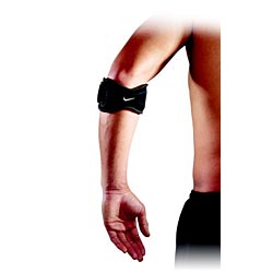
I find wrist immobilizers to be more effective than tennis elbow bands in the early phases of painful tennis elbow. The wrist splint puts the injured tendon at rest allowing the inflammation to calm down thereby relieving pain.
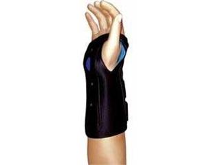
Other researchers found the tennis elbow band to be more effective. One study compared 3 common types of braces for their effect on grip strength in patients with lateral epicondylosis. In a randomized, controlled study of 52 patients, maximum and pain-free grip strength were assessed with the patient wearing an elbow strap orthosis, an elbow sleeve orthosis, a wrist splint, or a placebo orthosis. Use of the elbow strap and sleeve orthoses resulted in an immediate and equivalent increase in pain-free grip strength; consequently, the researchers suggest that either of these types of orthosis may be used. The wrist splint provided no immediate improvement in either pain-free or maximum grip strength.
Both corticosteroid and autologous blood injections have been shown to be effective. Corticosteroid injections at the lateral epicondyle have been shown to significantly decrease pain scores in the early post-injection period.
When the patient is free of pain through a full range of motion, begin strengthening therapy in a very slow and progressive way. When the patient regains strength and nears resumption of activity, place the emphasis on preventing future irritation (eg, correct technique or address equipment concerns in athletes who participate in racquet sports, modify jobs or activities in patients who are not athletes).
Therapy in patients who are having considerable pain from inflammation seems to be counter productive as it often aggravates the pain. Controlling inflammation and reducing pain enables patients to do more productive physical therapy.
The use of shockwave therapy raised initial excitement. However, 2 prospective, randomized, blinded trials showed no benefit of this intervention over placebo.
Surgical Therapy
A myriad of surgical procedures has been described for the treatment of lateral epicondylitis (tennis elbow). However, most surgical procedures involve debridement of the diseased tissue of the extensor carpi radialis brevis (ECRB) muscle with decortication of the lateral epicondyle. This procedure has been performed through open, percutaneous, and arthroscopic approaches.
Preoperative Details
Note the length of time of the patient’s symptoms. Also note the conservative therapeutic course that has been implemented, including any corticosteroid injections. Consider the patient’s workers’ compensation status, as patients with workers’ compensation claims may not respond as well to intervention.
A full evaluation should be performed on patients with lateral epicondylitis (tennis elbow) so that any other associated conditions can be detected.
Intraoperative Details
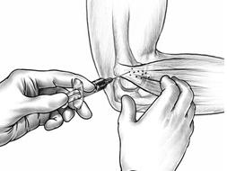
In the classic open-release procedure for lateral epicondylitis (tennis elbow), the patient is positioned supine. A 3-cm longitudinal incision is made over the lateral epicondyle. An incision is made through the extensor aponeurosis (outer elbow). The extensor carpi radialis brevis (ECRL) muscle is retracted medially, revealing the degenerative origin of the ECRB. All pathologic tissue is excised. The lateral epicondyle is decorticated with an osteotome, burr or by drill holes. The ECRL is sewn to the extensor aponeurosis in an attempt to repair the defect.

Lateral epicondylitis. Incision for open debridement of lateral epicondyle. Lateral epicondyle is circled.

Revision debridement for lateral epicondylitis. The fascia covering the origin of the extensor carpi radialis brevis muscle and the extensor carpi radialis longus muscle is fibrotic.

The extensor origin exposed

Lateral epicondylitis. Osteotome positioned over lateral epicondyle.
Postoperative Details
Surgical treatment of lateral epicondylitis (tennis elbow) is an outpatient surgical procedure. If the open approach is used, the elbow is usually protected initially with a splint or brace at 90°.
Follow-up
Early motion in a brace may be initiated at 2-3 days after surgical treatment of lateral epicondylitis (tennis elbow), with strengthening exercises usually started by 3 weeks, depending on the patient’s symptoms. Return to racquet sports can be expected by 4-6 months. Depending on the specific job requirements, patients can return to work in 6-12 weeks, although job modification or persistent use of a counterforce brace during work activities may be necessary.
I usually advise patients to wear the tennis elbow band for up to 6 months post op for heavy or demanding physical tasks to protect the healing tendon.
Complications
One of the most concerning complications of aggressive surgical debridement for lateral epicondylitis (tennis elbow) is lateral elbow instability. The proximity of the lateral collateral ligaments and the annular ligament makes them susceptible to injury. Other complications include recurrence or incomplete relief of pain.
Outcome and Prognosis
Surgical treatment of lateral epicondylitis (tennis elbow) has yielded predictably favorable results, with approximately 85% of patients reporting complete pain relief. Some patients may have persistent symptoms despite surgical treatment, and these patients may benefit from a more aggressive debridement.
Patients can link to my web site tennis elbow information here. You can download a booklet on lateral epicondylitis here and a booklet on medial epicondylitis here.
Thanks,
JTM, MD