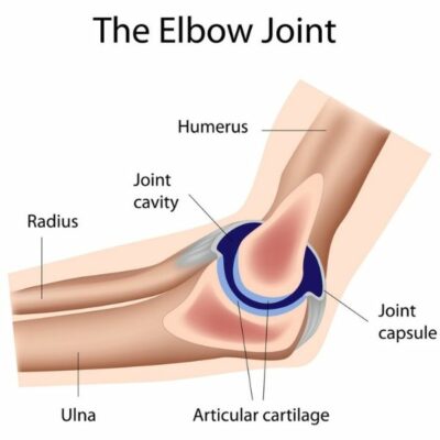
What is the anatomy of the elbow?
The elbow seems like a simple hinge; however, when the complexity of the elbow, forearm and wrist interaction are understood, it is easy to see why it can cause problems when not functioning correctly. Elbow injuries are common and can be extremely painful. Manchester, South Windsor, Enfield, Glastonbury and surrounding Hartford communities shoulder specialist, James Mazzara, MD is well trained and experienced in treating a number of elbow injuries.
The elbow joint works in unison with the shoulder and wrist to provide function and versatility to the arm. The versatile joint allows movement for flexion and extension, as well as rotation and twisting of the lower arm.
What are the important structures of the elbow?
The important structures that make up the anatomy of the elbow can be broken into several categories. These are:
- Bones – three bones make up the elbow joint;
- Humerus – long bone of the upper arm, connects to the shoulder joint and the elbow joint.
- Radius – one of the two bones in the forearm, is located on the side of the thumb.
- Ulna – the other bone located in the forearm, is located on the side of the pinky-finger.
- Ligaments – there are several flexible, strong ligaments that allow for stability and function of the elbow. These three help prevent injury;
- Annular ligament: The word annularmeans “ring shaped,” it forms a ring around the radial head and functions to keep the head of the radius in contact with the radial notch of the ulna.
- Radial collateral ligament (RCL): also called lateral collateral ligament (LCL) is responsible for extension of the joint and is located on the outside edge of the joint.
- Ulnar collateral ligament (UCL): also known as the medial collateral ligament, it is located on the inner edge of the joint. The UCL is
- Muscles, tendons and nerves – these make up the remainder of the anatomy of the elbow.
- Biceps and triceps muscles and tendons: The biceps tendon attaches the large biceps muscle on the front of the arm to the radius. It allows the elbow to bend with force. This tendon can be felt crossing the front crease of the elbow when tightening the biceps muscle. The triceps tendon connects the large triceps muscle on the back of the arm with the ulna. It allows the elbow to straighten with force, such as when a push-up is performed.
- Flexor and extensor muscle: responsible for wrist and finger movement.
- Nerves: All nerves traveling down the arm pass across the elbow joint and help make up the anatomy of the elbow. These nerves are called the radial nerve, ulnar nerve and median nerve. They are responsible for carrying signals back to the brain such as touch, pain and temperature.
- Blood Vessels – also traveling along the anatomy of the elbow joint are large blood vessels that supply the arm and the joint with blood. The largest, called the brachial artery, travels across the front crease of the elbow. The brachial artery splits into two branches, just below the elbow: they are called the ulnar artery and the radial artery.
What are common injuries of the elbow joint?
The complex anatomy make-up of the elbow allows it to be in use all day during work, leisure and sporting activities. Since the joint is placed under stress frequently, it is prone to injury. When injured, patients will experience pain, tenderness, swelling and loss of motion. Common injuries of the elbow include:
- Tennis Elbow
- UCL Injury
- Golfer’s Elbow
- Arthritis
- Biceps and triceps tendon rupture
To learn more about the anatomy of the elbow and the various causes of elbow pain, please contact the orthopedic offices of Dr. James Mazzara, orthopedic elbow specialist in Manchester, South Windsor, Enfield, Glastonbury and surrounding Hartford communities.
