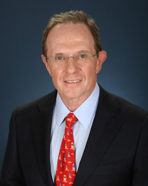
A medial plica is a remnant of an embryonic partition in the knee, and runs along the inner aspect of the kneecap.
It may also be called a ‘medial shelf’ – more a vertical shelf than a horizontal one, mind – and runs down from above the patella to the fat pad below the patella.
image of abnormal medial plica being nipped between femur and patella thickened medial plica photo taken at arthroscopy

illustration of medial and suprapatellar plica
As the knee bends the ‘shelf’ of tissue may also give a sudden snap over the rounded ends of the femur bone.

When a medial plica becomes abnormally thickened, it can be felt with the finger as a string-like object to the inner aspect of the kneecap.

Plica syndrome is often characterized by anterior knee pain, which is most commonly found along the superomedial aspect of the knee. The “plica” is due to remnant embryological tissue that compartmentalizes the knee during fetal development. The plica is considered a “vestigial” structure, which means that it has lost its ability to function over time and does not functionally affect an individual whether it is present or absent. It has been likened to the appendix, which can be a source of pain but lacks significant important function.

Symptoms


What are the symptoms of plica syndrome in athletes?

Usually plica syndrome is characterized by pain near the anteromedial (in front and toward the midline) side of patella (kneecap). Usually the pain is associated with bending of the knee and is irritated after and during exercise. Usually, no other symptoms other than pain is present, but it can occasionally be accompanied by swelling of the knee after prolonged physical activity.

How is plica syndrome diagnosed in athletes?

Plica syndrome is a difficult diagnosis in athletes, and is often made after “exclusion” of other common, structural causes of knee pain. Athletes will typically complain of discomfort along the front or inside of the knee. Usually there are no “mechanical symptoms” of locking or catching with a plica, but a sense of snapping or catching from a large plica can rarely occur. There may be tenderness in the region of the plica or medial femoral condyle where the plica is abrading the bone. Plain x-rays are usually normal. An MRI may show the plica on certain views, but is primarily useful to exclude bone bruises, meniscus tears, ligament injuries, and cartilage defects that can also cause similar pain and swelling of the knee joint. Routine labs are not beneficial to diagnose plica syndrome, but can help to identify other potential causes of knee pain.

Causes

What initially causes plica syndrome in athletes?

Most often plica syndrome is precipitated by a history of blunt trauma to the front of the knee or prolonged strenuous exercise. The plica is an embryologic remnant that is present throughout life, but is believed that a traumatic event or repetitive microtrauma is responsible for inciting inflammation and symptoms.

Treatment


What is the nonoperative treatment for plica syndrome in athletes?

Usually the first-line of treatment for plica syndrome in athletes includes rest from strenuous or precipitating activities. Physical therapy can be initiated to strengthen and stretch the muscles and soft tissues around the knee. In addition, oral non-steroidal anti-inflammatory medications and steroid injections can help alleviate the symptoms of plica syndrome by decreasing the inflammation and synovitis. If symptoms of pain and swelling completely resolve, a gradual rehabilitation program is started with a controlled return to competition. Steroid injections should be used judiciously, however. Occasionally, athletes may have a “flare” reaction after the injection with increased pain and swelling in the knee joint that resolves after 48 to 72 hours. Diabetics may have to monitor their blood sugar levels more closely due to a risk of hyperglycemia. In addition, there is a small risk of local “fat atrophy” and slight skin discoloration at the site of the injection.
What happens if conservative measures fail to help?

Failure to respond to months of nonoperative management may be an appropriate indication for surgical treatment in athletes. This procedure can be performed in a minimally invasive fashion (“arthroscopically”), using a camera and small instruments to excise the plica and associated inflammatory tissue around it that is causing the pain. The arthroscopic procedure also offers the benefit of allowing a thorough inspection of the knee for any other injuries or offending structures that may be a source of pain in athletes. The procedure has been reported with successful outcomes, allowing athletes to return to their previous level of competition without discomfort. However, the accurate diagnosis of a symptomatic plica remains the predominant challenge – not its surgical treatment. Many athletes will have asymptomatic plicas on imaging studies or arthroscopy that may not be the predominant cause of knee pain.
When can I return to play after surgery?

Arthroscopic surgery and plica excision is a relatively minor surgery. Unless associated procedures or repairs to knee ligaments or cartilage is required, a controlled rehabilitation program and quadriceps strengthening usually allows for an expeditious return to play 4 to 6 weeks after surgery.

Summary for the physician:

  1. Definitions (based on arthroscopy)
  2. Pleats of synovial lining of the knee
  3. Four Plicae
    1. Inferior plica (ligamentum mucosum)
      1. Intercondylar
      2. Anterior to anterior cruciate ligament
    2. Superior plica (plica synovialis suprapatellaris)
      1. Suprapatellar region (2 cm above patella)
    3. Medial plica (plica synovialis patellaris)
      1. Five anatomic subsets of the plica
      2. One finger breadth lateral to medial patella
    4. Lateral plica (rarely injured)
      1. Five anatomic subsets
  4. Mechanism of Injury
    1. More common in teenage athletes
    2. Onset often after blunt or twisting knee injury
    3. Repetitive knee flexion-extension
      1. Rowing
      2. Swimming
      3. Cycling
  5. Symptoms
    1. Medial patella pain
    2. Snapping sensation with knee flexion
    3. Provocative: Repeated flexion and extension motion
      1. Walking up and down stairs
      2. Squatting
  6. Signs
    1. Painful arc at 30 to 60 degrees
    2. Medial to patella above joint line
    3. Painful cord palpable alongside medial edge of patella
    4. Palpation reproduces symptoms
  7. Management
    1. Eliminate or reduce provocative activities
      1. Cycling
      2. Stair-step machine
    2. Local Cold Therapy
    3. Consider local Corticosteroid Injection
    4. Consider arthroscopic resection

    Thanks.

    JTM, MD